Fisiomonti | Fisioterapia | Roma Centro

admin

Clinic of the spine

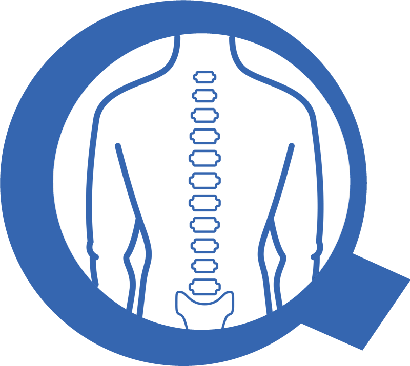
Clinic of the spine Vertebral column The vertebral column is composed of 33 vertebrae, subdivided into: 7 cervical vertebrae 12 thoracic vertebrae 5 lumbar vertebrae 5 calcified sacral vertebrae 4 calcified coccygeal vertebrae   The first three segments – cervical, thoracic, and lumbar – are characterized by the interposition of a disc between the vertebrae. The last two segments – sacral and coccygeal – have calcified vertebrae between them and are often considered as single bones. The function of the intervertebral discs is to stabilize the movement between the vertebrae and distribute the load. They are circular in shape and consist of two parts: • Nucleus pulposus: located in the center of the disc and has a semi-liquid consistency; • Annulus fibrosus: a fibrous ring located on the outer part of the disc that serves to protect the disc and contain the nucleus pulposus. Disc herniation and disc protrusion. Disc herniation is a condition in which, due to significant stresses, the nucleus pulposus protrudes from the annulus fibrosus.   It is necessary to distinguish disc protrusion from disc herniation: Disc protrusion: partial protrusion of the disc from its normal anatomical position Disc herniation: complete protrusion of the nucleus pulposus from the fibrous ring.   The disc segments most prone to developing herniation or protrusion are the first and last, respectively called L1-L2 and L4-L5 and L5-S1. This is because these vertebrae, being adjacent to the rigid dorsal tract and the sacrum (which is even more rigid), often move more than necessary to compensate for the reduced movement of these tracts. However, hernias or protrusions do not always cause pain. Disc herniation causes pain when it protrudes near the nerve root and consequently compresses it. In addition to pain, which can be local or even radiate sometimes to the fingers of hands and feet, other symptoms of disc herniation or protrusion include: • Tingling that can radiate along the territory innervated by the compressed nerve root; • Paresthesia and loss of strength that typically occur in more advanced conditions. Treatments for disc herniation and protrusion.   The remedies for lumbar hernias are varied and can be mainly divided into two groups: • Invasive remedies: surgery • Conservative remedies: physiotherapy Conservative Treatment: Physiotherapy for disc herniation or protrusion is currently the best tool for treating this condition. The physiotherapy cycle will aim to improve the movement of the entire spine. Initially, however, to reduce local and radiating pain, the following are used: Unloading postures; Specific manual techniques such as pumping and traction; High-tech physical means such as high-power laser, diathermy, hyperthermia, ultrasound. Once the pain is reduced, the recovery of correct posture is pursued. This goal is achieved with specific exercises, mobilizations, and small adjustments that the patient will have to adopt daily, such as the application of a lumbar cushion while working or driving. Sometimes, in conditions where there is particularly precarious muscle stability, the use of a lumbar belt during the day can be very helpful. In the final part of the therapeutic plan, efforts are made to improve trunk functionality with a specific rehabilitative training plan tailored to the individual being treated.   Surgical Treatment: Surgery for lumbar hernia involves the surgical removal of the hernia compressing the nerve root. It does not always achieve the desired result and leaves a fairly significant scar tissue, which is why it is performed only in severe cases where the hernia compression risks heavily damaging the nerve. BOOK NOW

Knee Clinic

Knee Clinic The knee is one of the major joints in the body where bones, cartilage, ligaments, muscles, and tendons work together in order to enabling a person to walk, run, jump, turn, and bend smoothly. The bones that make up the knee joint are 3: the femur, the tibia, and the patella. The femur and tibia form the femoro-tibial joint, which allows the movements of flexion and extension. The femur and patella, instead, form the femoro-patellar joint. A healthy knee has bones with very smooth surfaces covered by a tough protective tissue called cartilage. In addition to bones, the joint has ligaments positioned on the sides and inside of the knee that hold the joint’s bones in place and stabilize it during movement, together with muscles and tendons. Between the tibia and femur are two crescent-shaped fibrocartilaginous structures: the menisci, which protect the articular cartilage and act as shock absorbers. In areas of high friction, we find synovial bursae: sacs filled with fluid (called synovial fluid) that cushion the area where skin and tendons slide over the bone. Completing the joint is a joint capsule that lines it and secretes a lubricating fluid that further reduces friction and facilitates movement. Knee Pathologies Knee Osteoarthritis Knee osteoarthritis (or gonarthrosis) is a degenerative disease of the knee’s articular cartilage, leading to limited joint mobility. About 12% of the adult population and 90% of people over seventy suffer from knee osteoarthritis. Healthy articular cartilage protects the knee, cushioning it during movements and impacts. Cartilage naturally deteriorates due to the normal aging process. However, under certain circumstances, this wear and tear can become excessive and lead to pathologies. In fact the cartilage wears away progressively and becomes frayed and rough. The protective space between the bones decreases, and this can lead, in some cases, to the rubbing of the knee bones against each other. As this process progresses, the joint capsule thickens, thigh muscles become hypotonic and cause stiffness in the knee, which loses mobility, remaining curved and/or flexed. Ligaments can also be damaged, leading to a feeling of instability and giving way.   How it arises, what are the symptoms, and what can we do for knee osteoarthritis? Symptoms: Knee osteoarthritis symptoms often develop slowly and worsen over time. Signs and symptoms of knee osteoarthritis include: Pain. The knee is painful during or after movement. Stiffness. Upon waking or after a period of inactivity, the knee feels stiff. Laxity. The joint may feel soft to the touch. Loss of flexibility. The joint cannot move through its full range of motion. Grinding sensation. When using the knee, a “grinding” sensation may be felt during movements. Bone spurs. Extra bone pieces form around the affected joint, feeling like hard lumps. Swelling. Often caused by inflammation of the soft tissues around the joint. If we experience one or more of these symptoms for some time, WHAT CAN WE DO? Medical Examination: We must proceed to obtain a clear diagnosis by undergoing a medical examination. During this visit, the doctor will ask questions about the symptoms and medical history of the patient, conduct a physical examination in which he will check with various tests: tenderness, swelling, gait problems, clinical signs of muscle, ligamentous, or tendon injuries, and eventually order diagnostic tests, such as X-rays or blood tests. Diagnostic Tests: X-rays: Provide detailed images of dense structures, such as bone. They can help distinguish between various forms of osteoarthritis. X-rays of an arthritic knee may show narrowing of the joint space, changes in bone, and the formation of bone spurs. MRI: Occasionally, a magnetic resonance imaging or computed tomography scan may be necessary to determine the condition of the knee’s soft tissues. I have been diagnosed with knee osteoarthritis. What should I do? There are various possibilities: In the acute phase of the disease, one can start with analgesic treatment: The topical and systemic application of anti-inflammatory drugs can be effective in the acute phases of the disease (always consult your attending physician). As the acute phase subsides, various options open up (always subject to consultation and evaluation of the specific case with a trusted healthcare professional): Surgical Treatment: Arthroscopy or minimally invasive surgical treatment: this modern endoscopic technique can prevent the development of cartilage deterioration by removing damaged areas and pathological bone formation (osteophytes), as well as strengthening the ligament apparatus. Prostheses: Knee joint replacement surgery in case conservative and minimally invasive treatments have failed. Conservative Treatment (Physiotherapy): The main problems that the physiotherapist will have to face with a patient suffering from Knee osteoarthritis are: pain, loss of joint mobility, weakness, gait alterations and endurance. Considering that the damage to the cartilage cannot be reversed, one can reduce pain, improve mobility, function, and slow joint deterioration without surgical intervention but by working with the aim of: Strengthening the muscles surrounding the knee, glutes, and hip. Stretching tense and stiff muscles, such as the hamstrings. Encouraging fluid and nutrient exchange in the body with light aerobic exercises, such as walking, swimming, or pool exercises. Strong and flexible muscles will support the knee joint, resulting in less pressure on the damaged cartilage and bones. While excessive use of the knees can worsen joint health and knee osteoarthritis, on the other hand, the less the knees move, the weaker they tend to become. It is therefore necessary to find that balance to keep the knee joints moving just enough to keep them strong and healthy. In recent years, surgery has become a less used option for knee osteoarthritis. Evidence shows that physiotherapy alone is equally effective in relieving pain and improving knee joint condition in the presence of osteoarthritis.   MENISCAL INJURIES: The meniscus can be injured due to acute trauma or as a result of degenerative changes that occur over time. Meniscal injuries are often, but not exclusively, related to sports and usually occur during rotational movements: the athlete rotates or pivots the upper leg while the foot is planted, and the knee is bent. Sports

Tendinitis Clinic

Tendinitis Clinic In classical medical methodology, the diagnosis and study of the pathology underlying a symptom are always sought in medical treatment. However, this ideal model is not always applicable to tendons because: A) Pathologies related to symptomatic tendons have not  been clearly defined yet, and b) There is often a discrepancy between the reported/perceived pain and the evidence provided by diagnostic imaging, most likely due to the involvement and sensitization of the nervous system, which complicates the diagnosis.   The term “tendinopathy” itself does not indicate a specific pathology but it is a more generic term coined to indicate that “something is wrong” (a set of symptoms) in the tendon. Therefore, unlike diagnosing the pathology and the pain mechanism that cause the tendinopathy to occur, diagnosing “tendinopathy” is reasonably easy to achieve clinically, based on the perceived pain localized in the specific tendon when stressed. Tendinopathy is a debilitating problem that prevents individuals from leading an active life and indirectly impacts society.   The treatment of tendinopathies involves progressively loading and stressing the tendon. Activities that cause pain are initially completely excluded from treatment, followed by a slow and gradual introduction of exercises that stress the tendon below the pain threshold. For an effective treatment, patient collaboration is key, requiring adherence to a therapeutic exercise plan and avoidance of activities that improperly stress the tendon. The personalized therapeutic exercise plan will be monitored, updated, and implemented by the physiotherapist, who will simultaneously study motor patterns that may have contributed to the onset of tendinopathy.   In some types of tendinopathies, such as insertional tendinopathies (those affecting the tendon insertion, such as epicondylitis), performing high-load isometric and isotonic exercises (always below the pain threshold or performed in a position that does not cause pain) can even alleviate symptoms during the acute phase of inflammation. More generally, these types of exercises will be part of every rehabilitation plan, as they can help to manage components related to the nervous system and are crucial in reinstating the ability of the musculo-tendinous system to store and release energy before returning to more complex movements necessary for a return to sports practice.   Once the acute phase is overcome, corrective measures can be taken for the motor patterns that led to the onset of tendinopathy, using physical means such as taping, as well as additional therapeutic exercises aimed at correcting posture and execution of certain movements to prevent the recurrence of tendinopathy. Additionally, the therapeutic exercise plan may include targeted exercises to strengthen the muscles involved in the motor patterns associated with the initial onset of tendinopathy. BOOK NOW

×